3D Jaw Imaging
3D Jaw Imaging
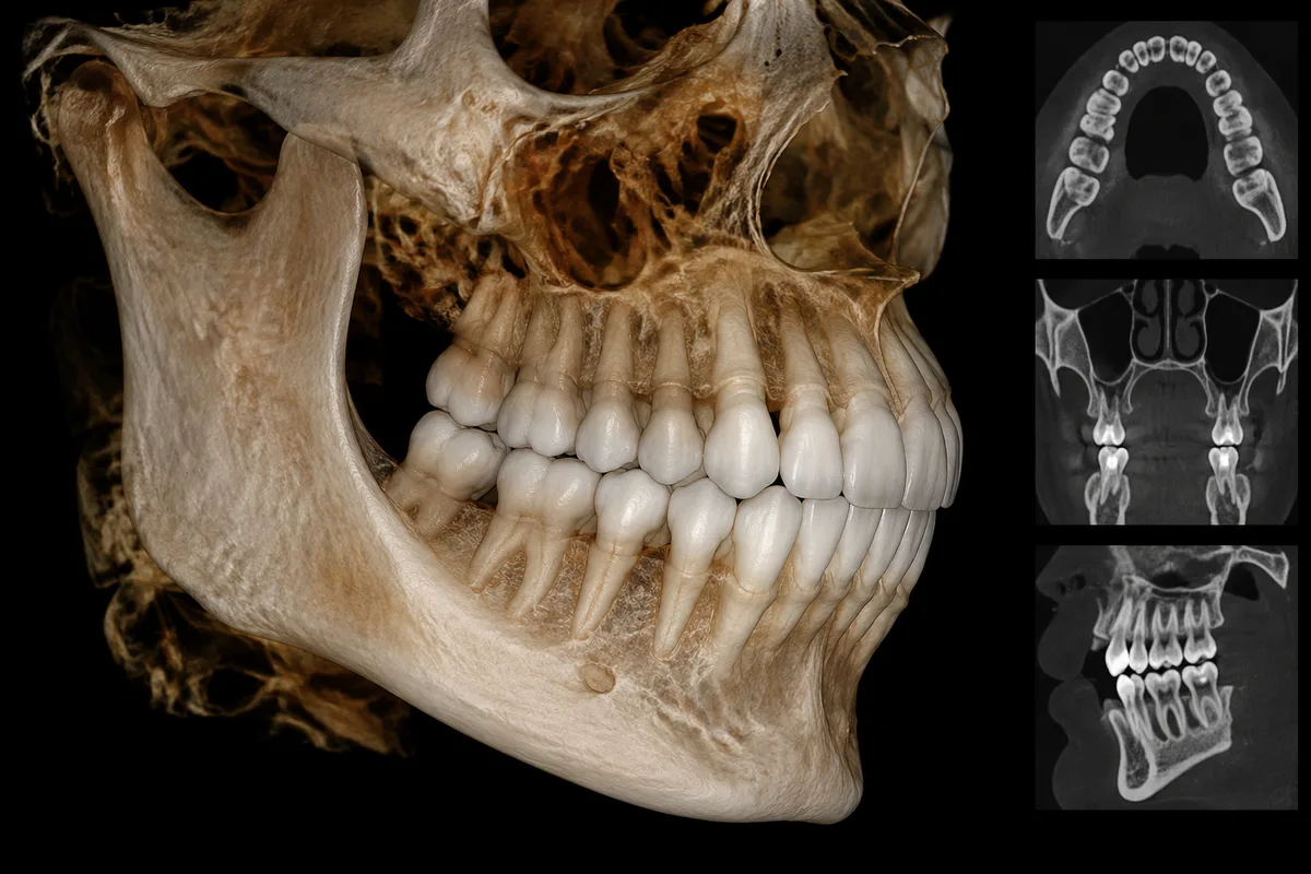
The practice offers on-site 3D CBCT (Cone Beam CT) imaging, giving Dr. Jeremy Montrose a detailed three-dimensional view of the jaw, joints, and airway. This level of detail supports careful, informed planning for TMJ, airway, and orthodontic care.
What 3D Jaw Imaging Shows
Unlike a flat two-dimensional X-ray, CBCT imaging captures the jaw, temporomandibular joints, and surrounding airway structures in three dimensions. This allows for a clearer picture of bone shape, joint position, and airway space than traditional imaging alone.
How It Supports Treatment Planning
- TMJ Evaluation — helps visualize joint structure as part of TMD evaluation and oral orthotic planning.
- Airway-Focused Dentistry — provides structural detail relevant to airway and sleep-disordered breathing evaluations.
- Orthodontic Planning — informs decisions around skeletal expansion, functional appliances, and orthopedic growth guidance for children and adults.
Who Oversees Imaging
Dr. Montrose reviews CBCT imaging as part of his broader physiologic approach to TMJ, airway, and orthodontic treatment planning, drawing on his training in airway-focused dentistry and functional orthodontics.
This content is for general information only and isn't dental or medical advice. Talk with a qualified provider about your own situation. If you have a medical emergency, call 911.