CBCT (Cone Beam CT) Imaging
CBCT (Cone Beam CT) Imaging
3D CBCT evaluation gives Dr. Montrose a detailed view of jaw, joint, and airway structure to guide TMJ, airway, and orthodontic treatment planning. For patients being evaluated for jaw joint discomfort, breathing concerns, or orthodontic needs, this imaging offers a clearer picture than flat, two-dimensional X-rays alone, helping shape a treatment plan that reflects the actual anatomy involved.
What CBCT (Cone Beam CT) Imaging is
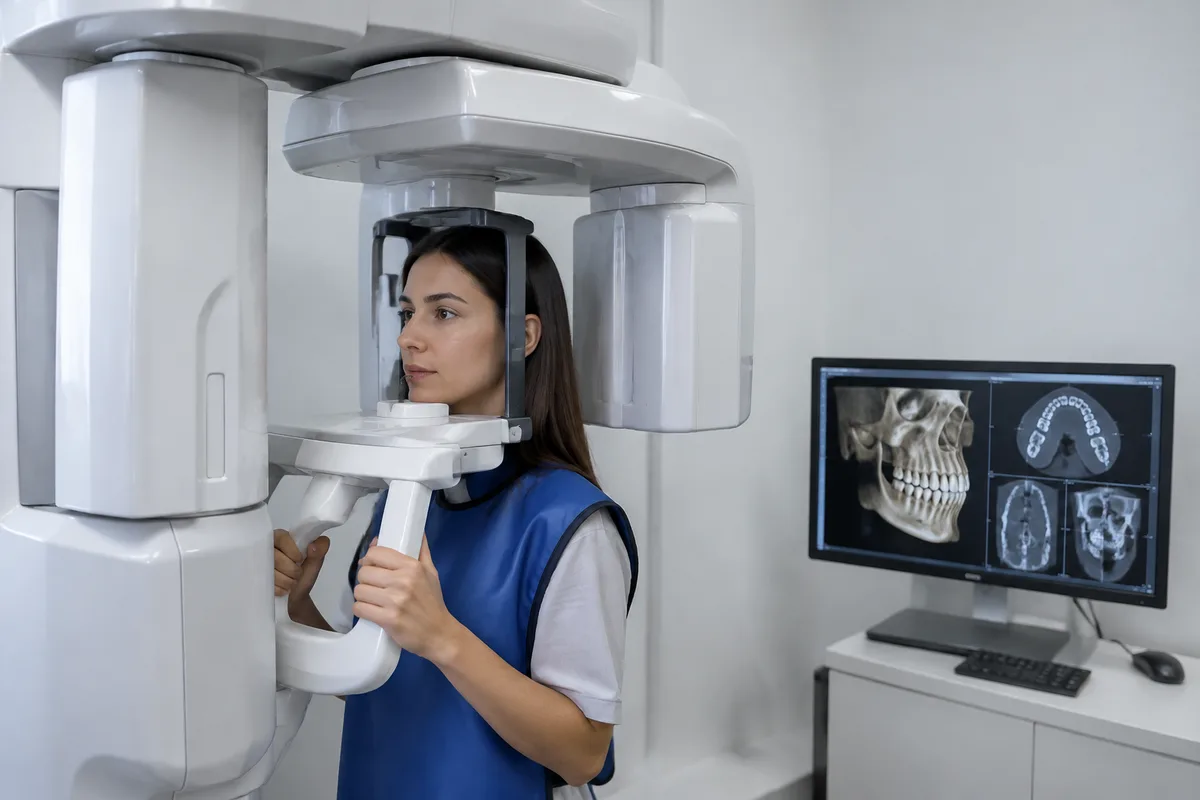
Cone Beam Computed Tomography, or CBCT, is a type of dental imaging that captures a three-dimensional volume of the jaws, teeth, sinuses, airway, and temporomandibular joints in a single scan. Instead of the flat, two-dimensional image produced by a traditional dental X-ray, a CBCT scanner rotates around the head and compiles many individual images into a detailed 3D dataset that can be viewed from any angle.
This technology improved on older imaging methods by giving dentists a way to see structures that overlap or hide behind one another on a standard X-ray, such as the joint surfaces of the jaw or the narrowing of the airway. Because CBCT units are designed specifically for the head and neck, the scan generally involves a lower radiation dose than a full medical CT, while still providing far more spatial detail than conventional dental X-rays.
How we use it at Jeremy Montrose, DMD
In the office, CBCT imaging is used to give Dr. Montrose a detailed, three-dimensional view of jaw, joint, and airway structure, which informs treatment planning for concerns such as TMJ evaluation and treatment, airway-focused dentistry evaluation, and sleep-disordered breathing evaluation. Because the scan captures bone and airway anatomy in a single volume, it can help clarify how the jaw joints are positioned, how the airway is shaped, and how the teeth and jaws relate to one another.
This imaging is also used alongside orthodontic planning, including early orthodontic intervention, skeletal expansion, and orthopedic growth and remodeling appliances, where understanding the underlying bone structure helps guide decisions about appliance design and treatment timing. CBCT works alongside the practice's digital X-ray technology, which is used for routine diagnostic imaging with reduced radiation exposure; together, these tools give Dr. Montrose different levels of detail depending on what a particular evaluation calls for.
What it means for you
A three-dimensional view can make it easier for a dentist to explain what is happening inside the jaw, joints, or airway, since a 3D model is often more intuitive to understand than a flat image. This can support clearer conversations about why a particular treatment approach, such as an oral orthotic, an expansion appliance, or a referral for further evaluation, is being recommended.
Because a single CBCT scan captures a large volume of anatomy at once, it can sometimes reduce the need for multiple separate X-rays taken from different angles. Modern CBCT units are also generally designed to limit radiation exposure relative to their diagnostic value, which is one reason this type of imaging is commonly used in dentistry today rather than reserved only for medical settings.
What to expect during your visit
Before the scan, you will typically be asked to remove any metal objects, such as earrings, glasses, or removable dental appliances, since metal can interfere with image quality. You will be positioned standing or seated, often with your chin resting on a support and a strap gently stabilizing your head to keep it still during the scan.
The scanner itself rotates around your head, usually taking well under a minute to complete its pass, though the full appointment time also includes positioning and any preparation beforehand. The scan itself is painless; you may hear a soft whirring sound as the machine moves, and you are simply asked to remain still and follow brief instructions, such as holding your breath for a few seconds if requested.
Afterward, there is no recovery time needed, and you can resume normal activities immediately. Dr. Montrose reviews the resulting images as part of your evaluation, using them to inform discussion of findings and next steps for your treatment plan.
Is CBCT imaging safe?
CBCT scanners are built specifically for imaging the head and neck and are designed to use a focused radiation dose appropriate for that purpose. As with any imaging that uses radiation, it is used when the information it provides is meaningful for diagnosis or treatment planning, rather than as a routine, unnecessary step.
Is the scan uncomfortable?
No, a CBCT scan is not painful. The main requirement is holding still for a short period while the scanner rotates around your head, which most patients find easy to manage.
How long does a CBCT scan take?
The actual scanning process is typically brief, often completed in under a minute, though your appointment will also include time for positioning and preparation. How often CBCT imaging is needed depends on your individual evaluation and treatment plan, as determined by Dr. Montrose.
Who is CBCT imaging appropriate for?
CBCT imaging can be useful for patients of various ages being evaluated for jaw joint concerns, airway and breathing issues, or orthodontic treatment planning, including growth-related evaluations in children. Whether it is appropriate for a particular patient depends on the specific concern being assessed, which Dr. Montrose will discuss as part of your evaluation.
If you would like to learn more about CBCT imaging or discuss whether it fits into your treatment plan, reach out to Jeremy Montrose, DMD to schedule a visit.
This content is for general information only and isn't dental or medical advice. Talk with a qualified provider about your own situation. If you have a medical emergency, call 911.